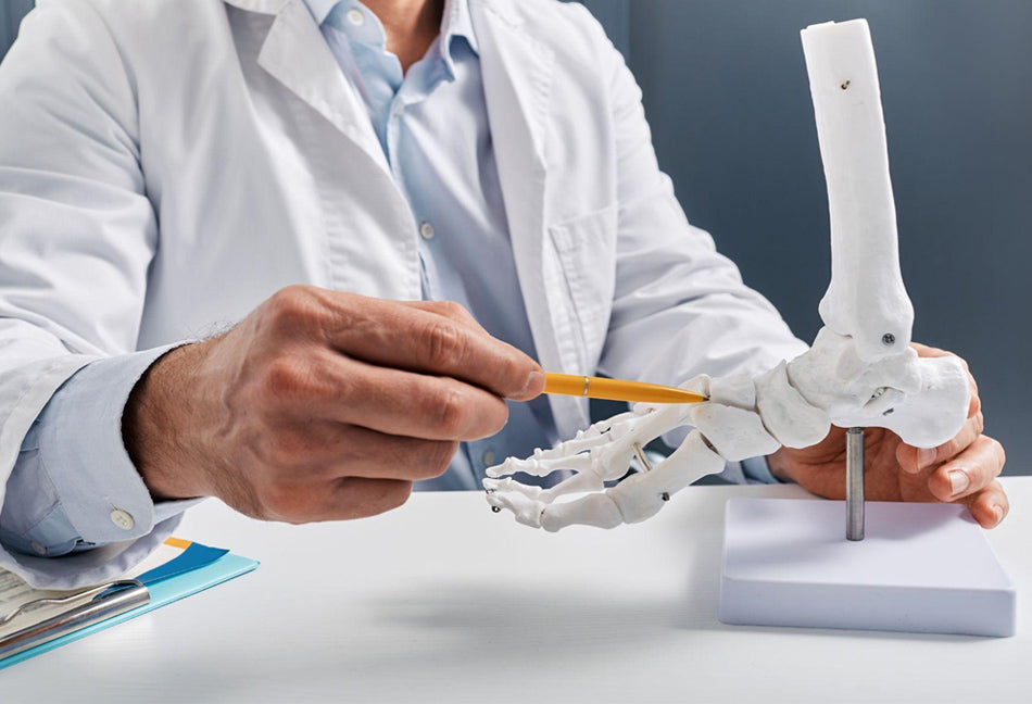
If you want to get serious about looking after your feet in the best way possible, then learning more about the different parts of the foot and how they work is a great place to start. Not only can you learn more about how certain foot problems develop – giving you the opportunity to take action sooner – you can also learn the right terms and phrases to use when discussing foot health with your podiatrist or other healthcare professional. This can make it much easier for you to understand any information or advice you’re given, allowing you to make informed decisions about your foot health.
So, without any further ado, let’s take a look at the bones of the foot.
How many bones are in the human foot?
If you’ve studied any anatomy before, you might be aware that the adult human body is made up of just over 200 bones in total – and these make up the familiar skeleton we recognise in doctors’ offices. That might sound like an awful lot of bones, but roughly 25% of those bones are located in the relatively small area of your feet.
In fact, each of your feet is made up of a whopping 26 bones, many of which are tiny compared to more well known bones such as the femur or the humerus. But those bones work together with muscles, tendons and ligaments to create the ease of movement enjoyed by a healthy foot – allowing us to walk, run and dance to our hearts’ content.
Within your feet, all those tiny bones are connected together with ligaments to keep them in the right form and position. Your bones are also connected to your muscles with the help of tendons, and it’s your muscles which contract and flex to create movement.
Sometimes, though, things can go wrong with this system of interconnected tissues and the bones can sit in different positions. This can affect the way your bones carry the weight of your body and put strain on other joints such as your knees or hips, causing pain. Everyone’s feet are unique, and what’s healthy for you might not be right for others. However, some foot positionings such as overpronation and supination can increase the risk of pain and inflammation. Fortunately, these issues can be corrected with the help of posture-adjusting insoles that encourage your feet to sit in a healthier position.
What are the bones in the foot called?
Since there are so many bones in the foot, we won’t go into the names of every single one of them. Instead, we’ll look at the different groups of bones within the three sections of the foot, as well as the roles they play to support movement and stability within the body – starting with the bones closest to the legs.
The Hindfoot
Although the term ‘hindfoot’ might sound a little odd, it’s just another name for the rear end of the foot where the heel meets the ankle. In fact, the heel is one of the main bones of the hindfoot region. Known to podiatrists as the calcaneus, the heel bone is the largest bone of the foot. As the part of the foot that lies directly below the knee and hip joints, the heel takes a lot of the strain needed to carry your body around.
Above the calcaneus, the talus bone is located on the dorsal, or top, part of the foot. While the calcaneus absorbs impacts from your foot striking the ground and carries your weight from the base of the foot, the talus then helps to spread that weight through to other bones chiefly located in the midfoot.
The Midfoot
The midfoot, as the name suggests, is located in the middle of your foot, covering the section between the heel and the ball areas. The first of the bones of the midfoot is the navicular bone, so-called because it resembles a boat. The navicular bone forms a joint with the talus behind it, and the cuboid bone to one side. The cuboid – which is, as you might have guessed, cuboid in shape – connects with the calcaneus behind it.
Then come the three cuneiform bones which form the transverse arch of the foot. This is the arch that goes from the left to the right of your foot, creating the bridge-like curve of the dorsal region. The cuneiforms – lateral, intermediate and medial – are wedge-shaped bones that attach the midfoot to the forefoot, as well as being the place where a number of muscles attach.
The Forefoot
The last section of the foot, known to podiatrists as the forefoot, is the front part which is made up of the ball of the foot and the toes. The first bones of the forefoot are the metatarsals, which form the ball of the foot. These bones can easily be damaged if you were to drop something heavy onto your foot because there isn’t a great deal of padding in the dorsal region. The metatarsal bones take your weight when you stand up on tip toes or wear high-heeled shoes.
Finally, the metatarsals lead into the phalanges, also known as the bones of the toes. Your four smaller toes on each foot have three phalanges. These are:
- The proximal phalanx – proximal meaning the closest to the rest of your foot
- The middle phalanx
- The distal phalanx – distal meaning the furthest away from the rest of your foot
Your big toes, on the other hand, only have two phalanges – the proximal and distal. Working together, your toes are integral for balance and stability, as well as allowing you to grip surfaces. Although we sometimes take them for granted, we’d be lost without our toes – which is why it’s so important to look after them and the rest of our feet.

